A incidência de Síndrome de Guillain-Barré (SGB) aumentou em cerca de cinco vezes no estado do Rio de Janeiro depois que o vírus Zika começou a circular no país. A estimativa é do neurologista Osvaldo Nascimento, pesquisador da Faculdade de Medicina da Universidade Federal Fluminense (UFF), que participa de estudo sobre fatores que, combinados com o Zika, ocasionam a SGB.
“A síndrome de Guillain-Barré continua sendo rara, só que nos chama atenção o fato de esses pacientes [que relatam ter passado por um quadro de Zika] apresentam o quadro clínico um pouco mais grave e com variantes da síndrome”, disse o especialista. Segundo Nascimento, antes, a média de ocorrência da SGB era 4 para cada 100 mil habitantes por ano, e agora fica entre 20 e 30 para cada 100 mil por ano.
A SGB é uma reação autoimune do organismo, que afeta os nervos periféricos e pode apresentar diferentes graus de manifestação, apresentando desde leve fraqueza muscular em alguns pacientes ao quadro raro de paralisia total dos quatro membros.
Em parceria com a Fundação Osvaldo Cruz (Fiocruz) e com outras universidades, a UFF está investigando qual o fator que, relacionado ao vírus Zika, pode desencadear a síndrome de Guillain-Barré. “Por que algumas pessoas têm a infecção aguda de zika e desenvolvem a síndrome, já que a grande maioria vai ter quadro agudo viral, com exantemas, as manchinhas vermelhas, conjuntivite, dor articular e depois passa?”, questionou Nascimento.
Em 2014, foram notificadas 1.439 internações por SGB em todo o Brasil no Sistema de Informação Hospitalar. Em 2015 , depois que o vírus Zika chegou ao país, o número chegou a 1.868 internações.
Alterações Neurológicas
Quadros de SGB e de encefalomielite, outra alteração no sistema imunológico, já foram associados também à infecção por dengue. Porém, segundo o neurologista, essa relação não impactou a incidência das duas doenças. “Você vê que nós tivemos muitos casos de dengue, mas, até agora, ninguém ouvia falar em Guillain-Barré”. Já o impacto das síndromes relacionadas ao vírus Zika foi evidente.
Encefalite e encefalomielite também são quadros neurológicos que podem ser desencadeados pelo vírus Zika. O quadro clássico de encefalite costuma começar com sonolência, instabilidade para andar, desorientação e pode acabar com o paciente precisando de aparelhos para respirar. Os quadros de encefalomielite acrescentam a estes sintomas dificuldades motoras. A intensidade dos sintomas varia muito. Também não se sabe quais fatores associados ao Zika podem fazer com que o paciente tenha essas alterações neurológicas.
O Ministério da Saúde diz que tem investigado as manifestações neurológicas identificadas em pacientes que estão em estados com circulação de infecções pelo Aedes aegypti, mosquito transmissor da dengue, do vírus Zika e da chikungunya. A pasta explica que entre essas síndromes se encontram encefalites, meningoencefalite, mielite e síndrome de Guillain-Barré, entre outras.
We describe the kinetics of Zika virus (ZIKV) detection in serum and urine samples of 6 patients. Urine samples were positive for ZIKV >10 days after onset of disease, which was a notably longer period than for serum samples. This finding supports the conclusion that urine samples are useful for diagnosis of ZIKV infections.
Zika virus (ZIKV) is an emerging mosquito-borne pathogen (family Flaviviridae, genus Flavivirus) that was isolated in 1947 from a rhesus monkey in the Zika forest in Uganda (1). ZIKV is believed to be transmitted to humans by infected Aedes spp. mosquitoes (2,3). Studies have demonstrated that ZIKV is endemic to Africa and Southeast Asia (4). Before 2007, few cases of human infection with ZIKV had been reported. In 2007, an epidemic of ZIKV infection in humans occurred in Yap, Federated States of Micronesia, in the Pacific region. A seroprevalence survey determined that ≤70% of the population had been infected (5). During 2007–2013, the few cases of infection with ZIKV reported were in travelers returning from Africa (6) or Southeast Asia (7).
In humans, ZIKV infection is characterized by mild fever (37.8°C–38.5°C); arthralgia, notably of small joints of hands and feet; myalgia, headache; retroorbital pain; conjunctivitis; and cutaneous maculopapular rash. ZIKV infection is believed to be asymptomatic or mildly symptomatic in most cases (5). Thus, Zika can be misdiagnosed during the acute (viremic) phase because of nonspecific influenza-like signs and symptoms. Hemorrhagic signs have not been reported in ZIKV-infected patients (5–7). However neurologic complications, including Guillain-Barré syndrome, have been observed (8).
Biological confirmation of ZIKV infections is based mostly on detection of virus RNA in serum by using reverse transcription PCR (RT-PCR). Although IgM against ZIKV can be detected by ELISA, few laboratories have this ability. Thus, in addition to the nonspecific clinical features of infection with ZIKV, laboratory diagnosis is challenging because of low viremia and cross-reactivity of ZIKV antibodies with other flaviviruses (including dengue), which require confirmation by neutralization assays (8) and make rapid serologic confirmation difficult. We investigated the diagnostic utility of urine as a source for detection of ZIKV RNA by real-time RT-PCR.
The Study
In October 2013, a ZIKV outbreak was reported in French Polynesia (9). This was the second outbreak of ZIKV infection reported in the Pacific region. In New Caledonia, where ZIKV infection had never been documented, the first cases of ZIKV infection imported from French Polynesia were confirmed by the end of November, and the first autochthonous cases were reported by mid-January 2014. Early in February 2014, the New Caledonia Health Authority declared an outbreak situation. By the end of August 2014, >1,400 cases of ZIKV infection were biologically confirmed, including 34 cases imported from French Polynesia (10).
Written informed consent was obtained from all patients in this study. Clinical signs and symptoms of 6 ZIKV-infected patients are shown in the Table. In this study, a cutaneous maculopapular rash of the trunk and extremities was systematically observed and considered a relevant clinical criterion. Complete blood counts showed a discreet perturbation common in many viral infections (mild leukopenia and thrombocytopenia associated with activated lymphocytes).
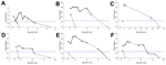
Figure. Detection of Zika virus in blood and urine specimens of 6 patients by using real-time reverse transcription PCR with primers/probe 1086/1162c/1107-Cy5 (11) New Caledonia, 2014. A) Patient 1; B)...
To detect ZIKV in samples (RNA extracted from 200 μL of serum or urine), we used both sets of primers/probe specific for ZIKV (11). A standard curve with serial dilutions of known concentrations of a ZIKV virus stock was used to estimate viral load in samples. All blood samples were also tested for dengue virus and chikungunya virus by real-time RT-PCR and showed negative results. ZIKV was detected in serum of 4 patients (Figure). Urine samples from 2 other patients were also positive for ZIKV, and showed a higher viral load than corresponding serum samples and were positive for ≤7 days (patient 4) and probably >20 days (patient 3) after viremia reached an undetectable level (Figure). Urine samples from 6 healthy patients were also assessed and showed negative results.
Partial sequences of the gene for ZIKV nonstructural protein were obtained (12) directly from amplification products from urine or serum samples. Sequences obtained (GenBank accession nos. KJ873160 and KJ873161) had 100% identity with the sequence of a ZIKV strain isolated from a patient who returned from French Polynesia in 2013. As observed previously (9,13), sequences also had 98% identity with sequences of ZIKV strains isolated in Cambodia in 2010 and in Yap in 2007.
Conclusions
We report the suitability of urine samples for diagnosis of ZIKV infection by showing that ZIKV RNA is detectable in urine at a higher load and with a longer duration than in serum. ZIKV infection has been poorly described because it is a benign, self-limiting illness in most cases (5). Thus, ZIKV infection has probably been underdiagnosed and underreported in disease-endemic settings (4) or in returning travelers. However, if perifocal vector control is to be implemented and severe neurologic complications are to be avoided, biological confirmation of ZIKV infection is essential. Because of the absence of specific IgM-based diagnostic tests, molecular confirmation is the only method available for routine diagnosis.
For ZIKV infection, date of onset of illness is difficult to establish because of sporadic and frequently mild fever. Although rash has been reported 3–5 days after the febrile phase (6,7), the 6 patients in our study had light asthenia and mild fever 2–3 days before the rash was observed; these symptoms were considered indicative of disease onset. Therefore, at the time the rash was observed, viremia was probably decreasing, which makes detection of virus in serum samples extremely challenging (Figure).
Other groups have reported that other flavivirus genomes, such as those of dengue virus (14), West Nile virus (15) and recently ZIKV (13), can be detected in urine samples for a longer time than in serum samples. Furthermore, use of urine samples for laboratory testing has some advantages, such as noninvasive sampling and ease of use. We detected ZIKV RNA in urine samples from all 6 patients. Urine samples showed strongly positive results; estimated maximum viral load was 0.7–220.106 copies/mL. For all cases with sequential specimens, ZIKV RNA was detected ≤15 days (range 10 days to >20 days) after onset of symptoms, which was >7 days after it was not detected in serum samples.
In our study, ZIKV was detected in patient serum until a rash was observed (days 2–3 after disease onset). However, urine was preferred for virus detection. We observed a slight increase in ZIKV RNA from urine over the first few days after disease onset and rash (Figure). We therefore attempted to isolate ZIKV from urine samples, but failed to cultivate infectious particles. Further investigations are needed to evaluate whether live infectious ZIKV particles are excreted in urine, as has been observed for other arboviruses (15).
This study investigated the diagnostic utility of urine as a source for detection of ZIKV RNA by real-time RT-PCR. Results suggest that urine might be useful for confirmation of ZIKV infection because virus was detected at higher titers and for a longer period in urine samples than in serum samples. Although these results need confirmation in larger cohorts, they strongly suggest the suitability of urine as a specimen for ZIKV detection and screening in large-scale investigations or other epidemiologic contexts (e.g., returning travelers).
In industrialized regions, where local transmission of arboviruses, such as dengue virus or chikungunya virus has been reported, physicians should test patients who return from tropical regions for ZIKV when a case of dengue-like infection is suspected. Travelers might be a source of local transmission because Ae. albopictus mosquitoes are a competent vector for ZIKV (3).
Dr. Gourinat is a clinical pathologist and head of the Serology, Immunology, and Molecular Biology Unit at the Institut Pasteur, Noumea, New Caledonia. Her research interests are diagnostic and biological surveillance of arboviruses.
Acknowledgment
We thank the staff of the Institut Pasteur in New Caledonia and M. Richard for providing technical support and I. Leparc-Goffart for providing Zika virus stock for the RT-PCR. Sequencing experiments were performed on La-Plateforme-du-Vivant (Noumea, New Caledonia).
References
Dick GW, Kitchen SF, Haddow AJ. Zika virus. I. Isolations and serological specificity.Trans R Soc Trop Med Hyg. 1952;46:509–20. DOIPubMed
Haddow AJ, Williams MC, Woodall JP, Simpson DI, Goma LK. Twelve isolations of Zika virus from Aedes (Stegomyia) africanus (Theobald) taken in and above a Uganda forest.Bull World Health Organ. 1964;31:57–69 .PubMed
Wong PS, Li MZ, Chong CS, Ng LC, Tan CH. Aedes (Stegomyia) albopictus (Skuse): a potential vector of Zika virus in Singapore.PLoS Negl Trop Dis.2013;7:e2348. DOIPubMed
Hayes EB. Zika virus outside Africa.Emerg Infect Dis. 2009;15:1347–50. DOIPubMed
Duffy MR, Chen TH, Hancock WT, Powers AM, Kool JL, Lanciotti RS, Zika virus outbreak on Yap Island, Federated States of Micronesia.N Engl J Med. 2009;360:2536–43. DOIPubMed
Kwong JC, Druce JD, Leder K. Zika virus infection acquired during brief travel to Indonesia.Am J Trop Med Hyg. 2013;89:516–7 and.DOIPubMed
Foy BD, Kobylinski KC, Chilson Foy JL, Blitvich BJ, Travassos da Rosa A, Haddow AD, Probable non-vector-borne transmission of Zika virus, Colorado, USA.Emerg Infect Dis. 2011;17:880–2. DOIPubMed
Oehler E, Watrin L, Larre P, Leparc-Goffart I, Lastere S, Valour F, Zika virus infection complicated by Guillain-Barré syndrome - case report, French Polynesia, December 2013.Euro Surveill. 2014;19:20720 .PubMed
Cao-Lormeau VM, Roche C, Teissier A, Robin E, Berry AL, Mallet HP, Zika virus, French Polynesia, South Pacific, 2013.Emerg Infect Dis.2014;20:1085–6. DOIPubMed
Lanciotti RS, Kosoy OL, Laven JJ, Velez JO, Lambert AJ, Johnson AJ, Genetic and serologic properties of Zika virus associated with an epidemic, Yap State, Micronesia, 2007.Emerg Infect Dis. 2008;14:1232–9. DOIPubMed
Kuno G, Chang GJ, Tsuchiya KR, Karabatsos N, Cropp CB. Phylogeny of the genus Flavivirus.J Virol. 1998;72:73–83 .PubMed
Kutsuna S, Kato Y, Takasaki T, Moi M, Kotaki A, Uemura H, Two cases of Zika fever imported from French Polynesia to Japan, December 2013 to January 2014.Euro Surveill. 2014;19:20683 .PubMed
Hirayama T, Mizuno Y, Takeshita N, Kotaki A, Tajima S, Omatsu T, Detection of dengue virus genome in urine by real-time reverse transcriptase PCR: a laboratory diagnostic method useful after disappearance of the genome in serum.J Clin Microbiol. 2012;50:2047–52. DOIPubMed
Barzon L, Pacenti M, Franchin E, Pagni S, Martello T, Cattai M, Excretion of West Nile virus in urine during acute infection.J Infect Dis.2013;208:1086–92 . DOIPubMed
Suggested citation for this article: Gourinat AC, O’Connor O, Calvez E, Goarant C, Dupont-Rouzeyrol M. Detection of Zika virus in urine. Emerg Infect Dis [Internet]. 2015 Jan [date cited]. http://dx.doi.org/10.3201/eid2101.140894
1These authors contributed equally to this article.
A SGB é uma reação autoimune do organismo, que afeta os nervos periféricos e pode apresentar diferentes graus de manifestação
A incidência de Síndrome de Guillain-Barré (SGB) aumentou em cerca de cinco vezes no estado do Rio de Janeiro depois que o vírus Zika começou a circular no país. A estimativa é do neurologista Osvaldo Nascimento, pesquisador da Faculdade de Medicina da Universidade Federal Fluminense (UFF), que participa de estudo sobre fatores que, combinados com o Zika, ocasionam a SGB.
“A síndrome de Guillain-Barré continua sendo rara, só que nos chama atenção o fato de esses pacientes [que relatam ter passado por um quadro de Zika] apresentam o quadro clínico um pouco mais grave e com variantes da síndrome”, disse o especialista. Segundo Nascimento, antes, a média de ocorrência da SGB era 4 para cada 100 mil habitantes por ano, e agora fica entre 20 e 30 para cada 100 mil por ano.
A SGB é uma reação autoimune do organismo, que afeta os nervos periféricos e pode apresentar diferentes graus de manifestação, apresentando desde leve fraqueza muscular em alguns pacientes ao quadro raro de paralisia total dos quatro membros.
Em parceria com a Fundação Osvaldo Cruz (Fiocruz) e com outras universidades, a UFF está investigando qual o fator que, relacionado ao vírus Zika, pode desencadear a síndrome de Guillain-Barré. “Por que algumas pessoas têm a infecção aguda de zika e desenvolvem a síndrome, já que a grande maioria vai ter quadro agudo viral, com exantemas, as manchinhas vermelhas, conjuntivite, dor articular e depois passa?”, questionou Nascimento.
Em 2014, foram notificadas 1.439 internações por SGB em todo o Brasil no Sistema de Informação Hospitalar. Em 2015 , depois que o vírus Zika chegou ao país, o número chegou a 1.868 internações.
Alterações Neurológicas
Quadros de SGB e de encefalomielite, outra alteração no sistema imunológico, já foram associados também à infecção por dengue. Porém, segundo o neurologista, essa relação não impactou a incidência das duas doenças. “Você vê que nós tivemos muitos casos de dengue, mas, até agora, ninguém ouvia falar em Guillain-Barré”. Já o impacto das síndromes relacionadas ao vírus Zika foi evidente.
Encefalite e encefalomielite também são quadros neurológicos que podem ser desencadeados pelo vírus Zika. O quadro clássico de encefalite costuma começar com sonolência, instabilidade para andar, desorientação e pode acabar com o paciente precisando de aparelhos para respirar. Os quadros de encefalomielite acrescentam a estes sintomas dificuldades motoras. A intensidade dos sintomas varia muito. Também não se sabe quais fatores associados ao Zika podem fazer com que o paciente tenha essas alterações neurológicas.
O Ministério da Saúde diz que tem investigado as manifestações neurológicas identificadas em pacientes que estão em estados com circulação de infecções pelo Aedes aegypti, mosquito transmissor da dengue, do vírus Zika e da chikungunya. A pasta explica que entre essas síndromes se encontram encefalites, meningoencefalite, mielite e síndrome de Guillain-Barré, entre outras.
O texto desenvolve a discussão acerca do aborto em casos de gestações envolvendo microcefalia e sua repercussão na sociedade.
Há exatamente setenta e cinco anos atrás, Adolf Hitler enviava carta ao seu General das SS, Reinhard Heydrich, determinando que este lhe apresentasse uma pronta solução final para a questão judaica. Heydrich respondeu afirmando que os nazistas deveriam destruir todos os judeus através de envenenamento, gás, fuzilamento, atos aleatórios de terror, doenças ou inanição, tudo em centros de morte consistentes em campos de extermínio estabelecidos no território polonês ocupado.
O saldo desse holocausto: mais de um milhão de crianças, dois milhões de mulheres e três milhões de homens judeus morreram nesses campos de horror. Anatoly Shapiro, o primeiro oficial do exército soviético a entrar no campo de concentração de Auschwitz, descreveu suas primeiras impressões sobre o que encontrou em 27 de janeiro de 1945:
“Não tínhamos a menor ideia da existência daquele campo. Nossos superiores não disseram coisa alguma sobre ele. Entramos ao amanhecer de 27 de janeiro. Havia um cheiro tão forte que era impossível aturar por mais de cinco minutos. Meus soldados não conseguiam suportá-lo e me imploraram para que fôssemos embora. Mas tínhamos uma missão a cumprir. Vimos algumas pessoas de pé em roupas listradas - eles não pareciam humanos. Eram pele e osso, somente esqueletos. Quando dissemos a eles que o Exército soviético os havia libertado, eles sequer reagiram. Não conseguiam falar ou mesmo mexer a cabeça. Os prisioneiros não tinham calçados. Seus pés estavam envoltos em trapos. Era janeiro e a neve estava começando a derreter. Até hoje não sei como conseguiram sobreviver. Quando chegamos ao primeiro pavilhão, estava escrito que era para mulheres. Entramos e vimos uma cena horrível. Mulheres desnudas e mortas jaziam perto da porta. Suas roupas tinham sido removidas pelas sobreviventes. Havia sangue e excrementos pelo chão. Nos alojamentos infantis, havia apenas duas crianças vivas. E elas começaram a gritar 'Não somos judias! Não somos judias'. Elas eram judias, mas estavam com medo de serem levadas para as câmaras de gás. Nossos médicos as tiraram dos alojamentos para serem limpas e alimentadas. Abrimos as cozinhas e preparamos refeições leves para os prisioneiros. Algumas das pessoas morreram porque seus estômagos não podiam mais funcionar normalmente. Vi os fornos e as máquinas de matar. As cinzas (dos mortos) eram espalhadas pelo vento”.
Ao final da guerra, a Assembleia Geral das Nações Unidas em Paris, aos 10 de dezembro de 1948, através da Resolução 217 A (III) elaborou a Declaração Universal dos Direitos Humanos, documento marco na história dos direitos humanos, para que nunca mais se repita esse trágico capítulo da história universal. Em um de seus considerandos, reza a Declaração: “O desprezo e o desrespeito pelos direitos do homem resultaram em atos bárbaros que ultrajaram a consciência da Humanidade.”.
Setenta e um anos após o holocausto dos judeus, lemos em revistas, jornais, periódicos, na internet e redes sociais que aviventam-se vozes propondo o livre direito ao aborto em gestações de bebês com microcefalia. E, mais do que isso, parece que a microcefalia reabriu a discussão sobre aborto no Brasil de forma generalizada.
Noutras palavras, vem-se propagando com muita força e ênfase uma solução final para os bebês com microcefalia. Em verdade, uma solução para a sempre inoperância e ineficiência do Poder Público no combate ao mosquito aedes aegypti, transmissor do vírus zika, este causador da microcefalia. E, também, uma solução para a falta de amor de alguns pais, disfarçada sob uma retórica nada convincente.
Aborto necessário, praticado por médico, se não há outro meio de salvar a vida da gestante é uma coisa, encontra previsão legal. Agora, aborto de bebês com microcefalia, não havendo risco à vida das gestantes, é genocídio.
Ora, o argumento de que a mulher não deve ser punida por uma falha das autoridades públicas em controlar o mosquito transmissor da doença não autoriza o exercício do direito de matar. Os bebês com microcefalia também são vítimas da falha do agente público. Aliás, em muito maior grau, pois só eles sentirão a doença na pele.
A gestante poderá até vir a renunciar à maternidade, através da chamada entrega consciente do bebê. Esclarece o Estatuto da Criança que “as gestantes ou mães que manifestem interesse em entregar seus filhos para adoção serão obrigatoriamente encaminhadas à Justiça da Infância e da Juventude” (§ Único, Art. 13). O dispositivo, naturalmente, também vale para os bebês com microcefalia. Entretanto, a gestante jamais terá o direito de decidir pela descontinuação dolosa de sua gravidez, optando pela morte de seu filho.
Não é por outra razão que o Código Civil, afinado com a Constituição Federal e os tratados de direitos humanos subscritos pelo Brasil, garante que a personalidade civil da pessoa começa com o nascimento com vida; mas a lei põe a salvo, desde a concepção, os direitos do nascituro.
A suprema ilusão do feto perfeito não pode e não deve permear as aspirações de uma Nação. Assiste inteira razão ao Apóstolo Paulo quando em suas cartas aos Coríntios diz que, sem amor, nada seria: “o amor é sofredor, é benigno; o amor não é invejoso; o amor não trata com leviandade, não se ensoberbece” (1 Coríntios 13:4).
O amor não mata!
Não se pode criar um ângulo ou um ponto de vista subjetivo sobre os direitos da pessoa humana. Ou todos os seres humanos, indistintamente, são dotados do sagrado e inalienável direito à vida, à liberdade e à igualdade; ou admitamos o triunfo do totalitarismo, da homogeneização da sociedade, da ideologia da superioridade racial.
Ao contrário do asseverado, a questão dos bebês com microcefalia não deve reabrir uma discussão sobre o aborto no País. Mas, sim, reafirmar o compromisso do Brasil em promover a dignidade da pessoa humana, a prevalência dos direitos humanos, o progresso da humanidade e, enfim, o bem de todos, sem preconceitos de origem, raça, sexo, cor, idade e quaisquer outras formas de discriminação.
↵* These authors contributed equally to this work.
↵‡ These authors contributed equally to this work.
↵† Present address: Vaccine Research Center, National Institute of Allergy and Infectious Diseases, Bethesda, MD 20892, USA.
Science 25 Feb 2016:
Abstract
Ebola virus disease in humans is highly lethal, with case fatality rates ranging from 25-90%. There is no licensed treatment or vaccine against the virus, underscoring the need for efficacious countermeasures. Here, we demonstrate that a human survivor of the 1995 Kikwit Ebola virus disease outbreak maintained circulating antibodies against the Ebola virus surface glycoprotein for more than a decade after infection. From this survivor we isolated monoclonal antibodies (mAb) that neutralize recent and previous outbreak variants of Ebola virus, and mediate antibody-dependent cell-mediated cytotoxicity in vitro. Strikingly, monotherapy with mAb114 protected macaques when given as late as five days after challenge. Treatment with a single human mAb suggests a simplified therapeutic strategy for human Ebola infection may be possible.
Ebola virus disease (EVD) causes severe illness characterized by rapid onset of fever, vomiting, diarrhea and bleeding diathesis (1, 2). The challenges of a large outbreak and the failure of traditional quarantine and contact tracing measures (3, 4) to control the 2014 West Africa outbreak highlights the urgency for therapies. The success in non-human primates (NHP) of ZMapp, a cocktail of three chimeric monoclonal antibodies (mAbs) derived from immunized mice (5–7), illustrated the potential of mAb therapies against EVD, and it is currently being evaluated in human trials. To date, efforts in NHP to simplify the ZMapp regimen to contain fewer mAbs have not been successful (7). We sought to isolate mAbs from human EVD survivors, with the goal of identifying antibodies that confer clinical protection either as single or dual-combination agents.
We obtained blood from two survivors of the 1995 Kikwit EVD outbreak (8) eleven years after infection. To determine if the subjects retained circulating antibodies against Ebola virus (EBOV) glycoprotein (GP), we assessed GP-specific antibodies by ELISA (Fig. 1A) (9). The reciprocal EC90 titer for subject 1 (S1) was 2,326, greater than ten-fold higher than control sera. Moreover, serum from the more severely ill subject, S1, displayed potent virus neutralizing activity (Fig. 1B), indicating that S1 maintained serologic memory against EBOV GP more than a decade following infection and suggesting the potential to clone immunoglobulins with potent neutralizing activity from S1’s memory B cells.
Fig. 1Isolation of antigen-specific monoclonal antibodies from Ebola virus disease survivor.
(A) Plasma obtained from two human survivors, an uninfected human donor and a non-human primate (NHP) vaccinated against EBOV GP were serially diluted and analyzed by GP ELISA, A450 (n = 1). (B) Lentivirus particles expressing luciferase and bearing EBOV GP were incubated in the presence of heat inactivated serum for 1 hour prior to addition to HEK293T. Infection was determined by measuring relative luminescence (RLU) after 3 days. Infection % = (RLU with serum / RLU without serum) X 100%, mean ± s.d. (n = 3, representative experiment shown). (C) Immortalized B cell supernatants isolated from Subject 1 were screened by EBOV GP ELISA A450 (n = 1). (D) Immortalized B cell supernatants from (C) were diluted 1:50, incubated with lentivirus particles and infection determined as in (B). Infection % = (RLU with supernatant / RLU without supernatant) X 100% (n = 1).
Therefore, we sorted memory B-cells from S1’s peripheral blood mononuclear cells, and immortalized individual clones with Epstein-Barr virus (10). Forty clone supernatants displayed a range of GP-binding (Fig. 1C), and two, 100 and 114, expressed antibodies with markedly higher neutralizing activity than all others (Fig. 1D). A second immortalization yielded 21 clones, from which the GP-specific clones 165 and 166 were rescued (fig. S1).
mAb100, mAb114, mAb165 and mAb166 sequences were PCR-amplified and antibodies produced by transient transfection. We assessed ELISA binding to EBOV GP and observed that mAb114, mAb165 and mAb166 displayed nearly 50% higher maximal binding than KZ52, a prototypic EBOV GP-specific human mAb (11), and 25% higher than 13C6, a component of the ZMapp cocktail (6, 7) (Fig. 2A). The binding curve of mAb100 plateaus similarly to KZ52 (Fig. 2A). mAb100 and mAb114 achieved half maximal binding (EC50) at a concentration of 0.02 μg/mL, which is 7-19 fold lower than the other mAbs. mAb165 and mAb166 had binding profiles similar to each other, with EC50s of 0.38 μg/mL and 0.35 μg/mL respectively, while EBOV control mAbs KZ52 and 13C6 had EC50s of 0.33 μg/mL and 0.14 μg/mL (Fig. 2A).
Fig. 2Characterization of purified EBOV GP monoclonal antibodies.
(A) EBOV GP ELISA in the presence of purified monoclonal antibodies as indicated, A450, mean ± s.d. n = 3, representative experiment shown) (B) Pseudotyped EBOV GP lentivirus particles were incubated with increasing amounts of purified monoclonal antibodies and infection measured as in Fig. 1B. Infection % = (RLU with antibody / RLU without antibody) X 100%, mean ± s.d. (n = 3, representative experiment shown). (C) V gene usage, sequence analysis and IgG subclass of antibodies from Subject 1. (D) Schematic of mAb100 and mAb114 UCA and variants created for investigation of the binding requirements of these mAbs. Shaded areas represent sequence from unmutated common ancestor (UCA) and light regions are from the somatic, mature antibody. Wild type, somatically mutated heavy (sH) or light (sL) chains; gH or gL, germline V-gene revertants of sH or sL in which the HCDR or LCDR3 are mature; gH-FR or gL-FR, germline V-gene revertants of sH or sL in which the HCDRs or LCDRs are mature; gH-FR1-2-4, germline V-gene revertants of sH in which the HCDRs and HFR3 are mature; gH-FR3, germline V-gene revertants of sH in which the HCDRs and HFR1, HFR2 and HFR4 are mature. (E and F) Binding to EBOV GP expressed on the surface of MDCK-SIAT cells by versions of mAb100 (E) and mAb114 (F) in which all or subsets of somatic mutations were reverted to the germline sequence. Shown is the ratio between the EC50 values of the variants and the wild-type (sH/sL). Ratio values above 100 indicate a lack of detectable binding (n = 1).
We next evaluated S1 mAbs capacity for neutralization using lentiviral particles pseudotyped with EBOV GP, a BSL-2 method that has been demonstrated to recapitulate wild type EBOV (BSL-4) results (12) (Fig. 2B and fig. S2A). mAb165 and mAb166 exhibited similar half-maximal inhibition (IC50) concentrations of 1.77 and 0.86 μg/ml, respectively. mAb100 and mAb114 were the most potent, with IC50s about one-log lower (0.06 and 0.09 μg/ml, respectively) than mAb165 and mAb166. Notably, all four of the isolated mAbs inhibited 100% of the virus unlike KZ52, which consistently displayed only 80-90% maximum inhibition, and 13C6, which neutralized < 20% at 10 μg/mL. Importantly, S1 mAbs also potently neutralized the 2014 West African Makona-variant (fig. S2B). Neutralization of wild type EBOV particles by each of the isolated antibodies was confirmed by plaque reduction assay (fig. S3).
Sequence analysis revealed that S1 mAbs displayed between 85-95% and 89-97% germline identity for heavy and light chains, respectively (Fig. 2C). Analyses of germline gene usage and V(D)J recombination indicate that they originate from different B cell lineages. The role of somatic hypermutations for the two most potent antibodies, mAb100 and mAb114, were analyzed using variants that were partially or completely reverted to the unmutated common ancestors (UCAs) (Fig. 2D and fig. S4, A and B). The fully reverted mAb100 (UCA/UCA), as well as a variant with a single change from the UCA VL (UCA/gL, A89T), recognized cells expressing GP with 2-4 fold weaker binding compared with the fully matured antibody (Fig. 2Eand fig. S4, A and B). When three HCDR3 mutations (A96V/V103Y/Y114S) were introduced in the reverted germline antibody (gH/UCA), binding was comparable to the fully matured mAb100 (sH/sL) suggesting that three mutations are sufficient to confer the binding observed for the fully matured mAb100. In the case of the mAb114, the fully reverted version (UCA/UCA) demonstrated negligible binding to GP (Fig. 2F and fig. S4, C and D). Introduction of two mutations (A96V and Y108S) in the HCDR3 of mAb114 germline (gH/UCA) was sufficient to confer an increase in binding, although still not to the degree seen with the fully matured mAb. Since these mutations are located at the base of the HCDR3 loop, they likely do not make direct contact with GP and thus may have a stabilizing effect on the whole HCDR3. The fully matured light chain and the two HCDR3 mutations (gH/sL) were sufficient to confer binding equivalent to the fully matured mAb (sH/sL). Importantly, the fully mutated light chain gene (UCA/sL), can partially compensate for a lack of somatic mutation in the heavy chain (Fig. 2F and fig. S4, C and D). The presence of additional mutations on either VH or VL is required to achieve the level of the fully matured mAb114 binding. These results suggest a rapid pathway of mAb114 affinity maturation through one or two somatic mutations, which became redundant as further mutations accumulated, a finding that is reminiscent of what was recently observed for the generation of broadly neutralizing influenza antibodies (13).
Since mAb100 and mAb114 were the most potently neutralizing antibodies, they were considered optimal candidates for further evaluation in NHP. In order to assess the potential for combination therapy, we wished to first rule out antagonistic binding to GP. We found that each antibody bound to GP in the presence of the other, suggesting that they recognize distinct regions on GP (Fig. 3A) and therefore could be used as combination immunotherapy to maximize efficacy (14). To define the regions targeted by mAb100 and mAb114 we employed biolayer interferometry to assess GP binding in competition with mAbs KZ52 and 13C6, which have epitopes in the GP base and glycan cap, respectively (15, 16). We found that mAb100 competes with KZ52 for binding at the base of GP, while mAb114 recognizes at least in part the glycan cap region, as demonstrated by competition with 13C6 (Fig. 3, B and C).
(A) Inhibition of binding of biotinylated mAb114 (left) and mAb100 (right) to GP-expressing MDCK-SIAT cells by pre-incubation with increasing amounts of homologous or heterologous unlabeled antibodies. Shown is the percentage binding of biotinylated antibody (n = 1). (B and C) Biolayer interferometry competitive binding assay to soluble EBOV GP using mAb100, mAb114, KZ52, 13C6 and isotype negative control. Biosensors were preloaded with GP followed by the competitor and analyte antibodies as indicated. Analyte binding curves (B) and quantitated % inhibition (C) are reported (n = 3, representative experiment shown). (D) Antibody-dependent cell-mediated cytotoxicity (ADCC) assay was determined at 31.6 ng/mL of mAb100, mAb114 (n = 3, representative experiment shown), control antibody or derivative antibodies with LALA mutations that abrogate Fc-mediated killing (n = 1). ADCC activity is shown as % killing, mean ± s.d.
Since some EBOV GP antibodies have been suggested to mediate antibody-dependent cell-mediated cytotoxicity (ADCC) (17) the ADCC activity of mAb100 and mAb114 were determined in a flow cytometric assay (Fig. 3D). We found that mAb100 and mAb114 mediated ADCC with maximal activity observed at a mAb concentration of 0.03 μg/ml. Target cell killing was mediated through Fc receptors since mAbs containing Fc LALA mutations (18) abrogated ADCC activity. Therefore, these mAbs have the potential to induce direct killing of infected cells in vivo, a key viral clearance mechanism.
The presence of potent neutralizing and ADCC activity, and the absence of cross competition, supported testing mAb100 and mAb114 in vivo protective efficacy. We challenged four rhesus macaques with a lethal dose of EBOV. One day post-challenge, the treatment group (n = 3) received an intravenous injection with a mixture of mAb100 and mAb114 at a combined dose of 50 mg/kg, and the treatment was repeated twice at 24-hour intervals. Circulating GP-specific antibody titers in mAb recipients peaked after the second injection, and reciprocal ELISA titers remained above 105 throughout the study, suggesting minimal clearance of the mAbs (Fig. 4A). The untreated macaque succumbed to EVD on day 10 with a circulating viral load exceeding 108 ge/ml (Fig. 4, B and C). In contrast, all three mAb-treated macaques survived challenge without detectable viremia. Consistent with historic controls, the untreated animal displayed hallmark indicators of EVD including thrombocytopenia and elevations in alanine transaminase and creatinine from day 6 through the time of death (Fig. 4D and figs. S5 to S8). In contrast, the treatment group remained within normal ranges for these parameters, and free of all EVD symptoms.
Animals were challenged with a lethal dose of EBOV GP on Day 0 and given injections of antibody totaling 50 mg/kg at 24, 48 and 72 hours post-exposure (A to H) or at 120, 144 and 168 hours post-exposure (I). Surviving animals were euthanized at the conclusion of the study (Day 28). (A) to (D): Challenge data from monoclonal antibody mAb114/mAb100 mixture. mAb114 monotherapy treatment beginning 24 hours [(E) to (H)] or 120 hours (I) after exposure. Treatment animals in black, untreated controls in red. (A) and (E): Ebola GP specific ELISA titer (reciprocal EC90). (B) and (F): Viremia in blood by qRT-PCR expressed as genome equivalents (ge) per mL. (C), (G) and (I): Survival. (D) and (H): Selected hematologic and chemistry data. Platelets (PLT), alanine transaminase (ALT), creatinine (CRE).
We next asked whether monotherapy is sufficient for protection, and focused on mAb114 since it showed higher maximal binding than mAb100. We exposed four macaques to a lethal dose of EBOV and administered 50 mg/kg of mAb114 (n = 3) to the treatment group after a one-day delay, followed by two doses at 24-hour intervals. All treated macaques survived, whereas the control animal succumbed to EVD on day 6 with a peak viral load of 1010 ge/ml (Fig. 4, E to G). In contrast to the previous experiment, transient viremia was observed in the treated animals (Fig. 4F), but remained at levels less than 0.1% of the untreated control animal. Despite transient viremia, treated animals remained free of clinical and laboratory abnormalities (Fig. 4H and figs. S9 to S12).
Since a delay in treatment is a distinct possibility in an outbreak setting, we evaluated the therapeutic potential for mAb114 when treatment was delayed until 5 days after lethal EBOV challenge. All three animals in the treatment group survived, while the control animal succumbed to EVD on day 9. Moreover, animals in the treatment group remained symptom-free and protected against thrombocytopenia, transaminitis and renal dysfunction (Fig. 4Iand figs. S13 to S16).
mAb114 has several characteristics that may contribute to protection as a monotherapy compared to KZ52 and 13C6, which were non-protective in NHPs (7, 19). First, while KZ52 and mAb114 potently neutralize EBOV in vitro, only mAb114 completely neutralizes input virus. Secondly, mAb114 does not require complement for neutralizing activity (Fig. 2B) in contrast to 13C6 (6). Based on these observations, one hypothesis is that protective monotherapy may require both potent binding and complete complement-independent neutralization. In addition, mAb114’s specific mechanism of neutralization, which targets an essential step in virus entry (20), and the observed in vitro ADCC activity may contribute to mAb114’s ability to protect against lethal EVD in macaques.
In these studies, we showed that antibodies as well as memory B cells specific to EBOV are maintained in a survivor more than a decade following infection. For the mAbs isolated from this survivor, a potential role for antibody-dependent killing is suggested by in vitro ADCC activity that in vivo may be mediated by multiple effector cells such as natural killer cells, macrophages or neutrophils. In addition, these mAbs showed potent neutralizing activity against Ebola GP variants that have evolved over a 40-year period. Together, these data demonstrate the therapeutic potential of these mAbs as dual- and/or mono-therapy and contribute to understanding the mechanisms of antibody-mediated protection against Ebola virus disease.
, Ebola virus disease in West Africa—the first 9 months of the epidemic and forward projections. N. Engl. J. Med. 371, 1481–1495 (2014).doi:10.1056/NEJMoa1411100pmid:25244186
, Ebola outbreak in Kikwit, Democratic Republic of the Congo: Discovery and control measures. J. Infect. Dis. 179 (suppl. 1), S259–S262 (1999).doi:10.1086/514302pmid:9988192
, An efficient method to make human monoclonal antibodies from memory B cells: Potent neutralization of SARS coronavirus. Nat. Med. 10, 871–875 (2004).doi:10.1038/nm1080pmid:15247913
,Rapid development of broadly influenza neutralizing antibodies through redundant mutations. Nature516, 418–422 (2014).doi:10.1038/nature13764pmid:25296253
, Structure of the Ebola virus glycoprotein bound to an antibody from a human survivor. Nature 454, 177–182 (2008).doi:10.1038/nature07082pmid:18615077
, Structures of protective antibodies reveal sites of vulnerability on Ebola virus. Proc. Natl. Acad. Sci. U.S.A. 111, 17182–17187 (2014).doi:10.1073/pnas.1414164111pmid:25404321
, Effector function activities of a panel of mutants of a broadly neutralizing antibody against human immunodeficiency virus type 1. J. Virol. 75, 12161–12168 (2001).doi:10.1128/JVI.75.24.12161-12168.2001pmid:11711607
, Neutralizing antibody fails to impact the course of Ebola virus infection in monkeys. PLOS Pathog. 3, e9 (2007).doi:10.1371/journal.ppat.0030009pmid:17238286
, Efficient generation of monoclonal antibodies from single human B cells by single cell RT-PCR and expression vector cloning. J. Immunol. Methods 329, 112–124 (2008).doi:10.1016/j.jim.2007.09.017pmid:17996249
, Endoproteolytic processing of the Ebola virus envelope glycoprotein: Cleavage is not required for function. J. Virol. 73, 1419–1426 (1999).pmid:9882347
, Transcriptional profiling of the circulating immune response to lassa virus in an aerosol model of exposure. PLOS Negl. Trop. Dis. 7, e2171 (2013).pmid:23638192
Acknowledgments: We thank M. Cichanowski for graphics, B. Hartman for manuscript preparation and A. Tislerics and J. McLellan for critical reading of the manuscript. We would like to thank the study volunteers for the donation of blood for these investigations. The data reported in this manuscript are tabulated in the main paper and in the supplementary materials. Antibody sequences were deposited to GenBank, accession numbers: KU594601, KU594602, KU594603 and KU594604. This work was supported by the Intramural Research Program of the Vaccine Research Center, National Institute of Allergy and Infectious Disease, and the National Institutes of Health. Nancy Sullivan, Sabue Mulangu, Barney Graham, Julie Ledgerwood, Daphne Stanley, Jean-Jacques Muyembe-Tamfun, John Trefry, Davide Corti, and Antonio Lanzavecchia are listed as inventors on patent applications related to anti-Ebola virus antibodies and their use. Opinions, interpretations, conclusions and recommendations are those of the authors and are not necessarily endorsed by the U.S. Department of Defense or the U.S. Department of the Army.


